Risk Factors:
As previously mentioned, people involved in strenuous physical activities such as athletes especially runners, dancers, or those who need to walk or stand for long periods are at greater risk.
Other factors enhancing the risk of plantar fasciitis include:
- Faulty foot biomechanics. Excessive inward rolling of the foot when walking puts extra stress on the ligament. As the foot flattens out on the ground, the ligament is stretched excessively. A repeated motion eventually damages the ligament.
- Abnormal arch shape, either high or flat
- Tightness in the calf muscles or Achilles tendon (tendon that attaches the calf muscles to the heel), leads to excessive pronation of the foot, which in turn stretches the fascia beyond its capacity, inducing damage.
- The incidence is higher between 40 to 60 years of age. Concurrent diabetes may also exacerbate the problem.
- Improper footwear such as loose, worn out or ill-fitting shoes that do not support the arch properly.
- Obesity or sudden weight gain as in pregnancy
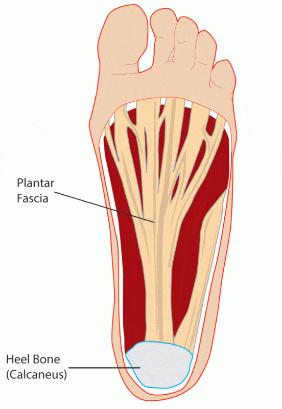
Heel spur, a small bony projection at the anterior aspect of heel bone, is often thought to be a cause of plantar fasciitis; however, new evidence suggests that these spurs are, in fact, the result rather than the cause of inflammation.
Diagnosis:
The location and nature of the pain as well as stiffness of the arch of the foot is quite characteristic of the condition. The bottom of the heel is tender to the touch. Pain worsens when the ankle is dorsiflexed, as it stretches the fascia.
X-rays or an MRI may be recommended in order to rule out other problems such as stress fractures or nerve entrapment. A heel spur may also be detected on radiological examination; however, as mentioned previously, its presence is not related to the pain.
Treatment:
The first line of treatment includes rest and supportive therapy.
- Get some rest (delayed treatment can worsen the condition and prolong the treatment duration).
- Apply ice to the painful area (effective in the first 24-48 hours)
- Use non-steroidal anti-inflammatory drugs to relieve pain, e.g. ibuprofen, naproxen etc.
- Foot massage
- Proper footwear, with well-cushioned sole and adequate heel support.
Innersoles:
Orthotic insoles such as arch supports, heel cups, felt pads etc., have been shown to improve symptoms. Both over-the-counter and custom-made innersoles have been found to be equally effective in relieving the pain of acute plantar fasciitis.
The basic aim of using innersoles is to provide support to the heel and distribute the load evenly to prevent excessive stretching of the already damaged fascia. This prevents further deterioration and aids in healing. innersoles also help by countering the contributing factors in plantar fasciitis such as over pronation, postural faults, a flat arch, etc.
Night Splints:
As the name indicates these are braces worn at night over the foot and lower leg. These splints keep the plantar fascia and the Achilles tendon in a mildly stretched position overnight. This helps to reduce the pain felt when taking one’s first steps in the morning.
There is some evidence that taping the foot helps as well. Taping is applied across the sole; it supports the arch of the foot and reduces stress on the fascia.
Physiotherapy:
Exercises involving stretching the calf muscles and plantar fascia help in reducing stiffness and pain in the fascia. These exercises have shown to improve symptoms for several months.
Steroids:
In more resistant cases steroids are usually injected, with or without needles. The needleless injection technique, iontophoresis, involves transdermal delivery of a drug through active transportation in an electric field. A steroid solution is applied on the affected area, followed by application of a small electric charge, mild enough not to produce any discomfort. However, repeated steroid injections may lead to weakening and subsequent rupture of the plantar fascia. Plantar fat pad atrophy has also been attributed to injecting steroids in the area. Moreover, as new research reveals that plantar fasciitis is a result of degenerative changes rather than inflammation, the role of steroids in its management is therefore questionable.
Extracorporeal Shock Wave Therapy (ESWT):
This involves focused delivery of low or high-energy shock waves to the body. These result in micro trauma that in turn initiates a healing response, which eventually repairs the damaged ligament.
Surgery:
Surgery carries its own risks and should only be considered as a last resort. Part of the plantar fascia is cut in order to release tension. This also helps decompress the inflamed flexor digitorum brevis muscle by providing more space, thereby relieving pain.
Less invasive surgical procedures such as ultrasound-guided needle fasciotomy (the fascia is released by inserting a needle) and coblation surgery (Topaz procedure) have also been used successfully in the treatment of resistant cases.
Immediate attention to the problem, as well as complete eradication of the causative factor, is mandatory for satisfactory treatment. Ignoring the problem may cause compensatory changes in the gait and posture resulting in knee, hip or lower back pain.