Plantar Fascia Anatomy
The word fascia comes from Latin, meaning “a band”. It denotes the layer of fibrous connective tissue that surrounds different organs, muscles, bones, blood vessels and nerves.
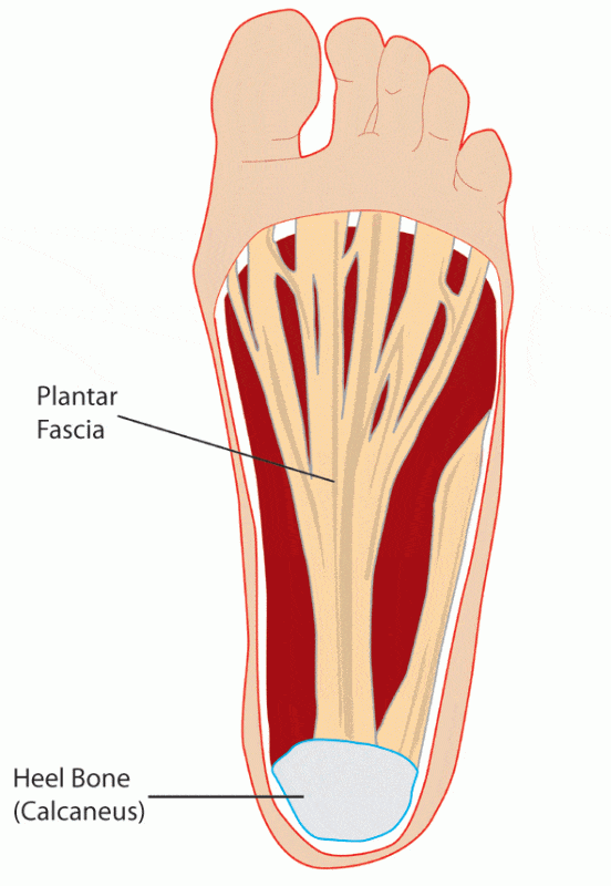
The plantar fascia is the fibrous tissue layer on the plantar surface of the foot that connects the heel bone to the toes. It supports the arch of the foot and protects the sole from injuries.
It is a thick white band of longitudinally extended collagen fibers. Some categorize it as fascia while others consider it an aponeurosis. An aponeurosis is defined as a white tendinous extension of a muscle, connecting it to the structures that it moves.
Attachments of Plantar fascia:
It originates posteriorly from the medial process of the tuberosity of calcaneus (heel bone). As it extends towards the toes, it divides into five separate bands that extend to the proximal phalanges (bones of the toes, closest to the foot).
The superficial layer of plantar fascia embeds into the skin fold between sole and the toes.
The deeper layer extends to embrace the digital flexor tendons on each proximal phalanx.